You are here
Researchers find that sugar is the solution for looking at platelets

Researchers at The Australian National University (ANU) have found a way to look through the walls of microchambers that are used to study thrombus (blood clot) formation using light sheet microscopy.
Platelets are the tiny cells that help form blood clots, which are imperative to stop bleeding, and are difficult to investigate under light sheet microscopy.
Light sheet microscopy is a powerful tool in biological imaging because live samples can be imaged quickly, without the fear of phototoxicity. But light sheet microscopes are incompatible with the microchamber used to study tiny platelets.
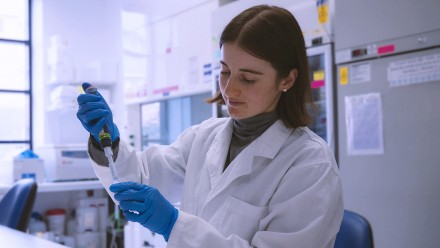
“When we use the light sheet microscope to look through the walls of the chamber, the platelets look distorted and blurry,” said Mr Tienan Xu, PhD candidate and lead author of the research.
The researchers found that by submerging the chamber in a solution of sucrose, table sugar you can buy at your local supermarket, the walls become invisible when viewed through the microscope, and the images of the platelets become clear.
“This simple imaging protocol will have big implications for future microchamber volumetric bioimaging,” said Dr Steve Lee, senior author of the research published in the journal Lab on a Chip.

Comparison of the chamber with (right) and without (left) the sucrose solution.
“Without the sucrose, looking through the walls of the chamber is like how a straight straw in a glass cup of water looks bent,” said Dr Daniel Lim, a postdoctoral researcher of the Lee Group and co-author of the research.
“It is the same when trying to look at platelets in the chamber - they look distorted.”
“Because blood flow is a fast and dynamic process, a distorted image makes the movement of platelets, and blood clot formation, very difficult to study.”
“The simple solution of submerging the microchamber in sucrose allows us to watch blood flow in real time using microscopes we already have.”

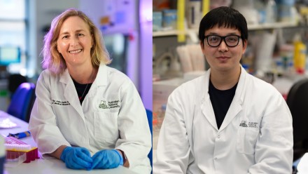
Ms Yujie Zheng, PhD Researcher and co-author of the research in Lab on Chip.
“Biologists have a different mindset to engineers when approaching problems,” said Yujie Zheng, PhD Candidate of the Lee Group.
“Biologists and blood researchers want to see clearer images of platelets using the microscopes they already have, and as engineers we are able to make this possible.”
“This application to medical research highlights the significance of the tools we have developed.”
“There is a perception that scientists moving from their undergraduate studies to postgraduate research should stick to their background training, but this is not the case, especially when there is an opportunity for collaboration,” said Dr Lim, whose research spans across the Colleges of Engineering and Health and Medicine.
“This type of collaboration broadens our understanding of scientific research and allows us to experience two different types of joy as scientists.”
“First, uncovering the hidden workings of a biological process such as blood clot formation, and the second, designing and creating a simple engineering solution that enables this type of research to happen.”

Dr Daniel Lim, researcher at the College of Health and Medicine and the College of Engineering and Computer Science.
Researchers at The National Platelet Research and Referral Centre (NPRC) can use this new tool to improve our understanding of platelet disorders.
“By studying the formation of blood clots, we get insight into conditions where platelet function is reduced and the patient is at risk of bleeding,” said Professor Elizabeth Gardiner, Head of the Cancer Department at JCSMR.
Tienan Xu and Yujie Zheng are PhD candidates at the Research School of Electrical, Energy and Materials Engineering. They conducted this research in the Imaging & Cytometry Facility at Australia’s national medical research institute and in collaboration with scientists from EMBL-UNSW Node and the ARC Centre of Excellence for Advanced Molecular Imaging. This work also received support by grants from the Australian Research Council, ANU Major Equipment Grant and the ARC Centre of Excellence Translational Photosynthesis.